


SpectraMax MiniMax 300 Imaging Cytometer
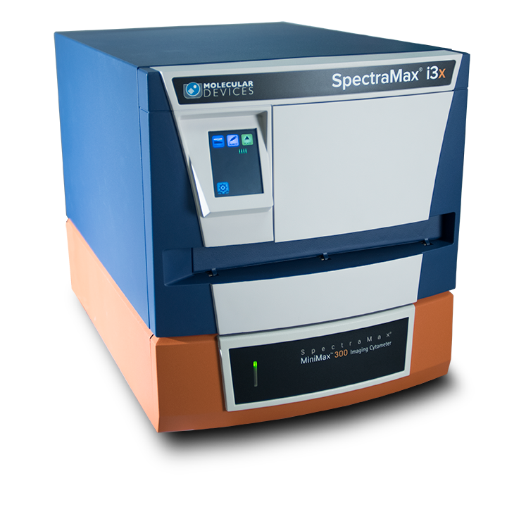
The SpectraMax MiniMax 300 Imaging Cytometer option is a field-upgradeable option for the SpectraMax i3/i3x Multi-Mode Detection Platform that offers cellular imaging capability.
- Simplified workflow: Add cellular imaging assays in a small footprint
- No Stain Analysis: Eliminate cell staining for cell counting and confluence measurements using StainFree™ Technology
- More data: Multi-parametric cell-based assays with brightfield and green and red fluorescent imaging
- Familiar software: Set up, image, and analyze cells in a snap using SoftMax® Pro Microplate Data Acquisition & Analysis Software
- Higher sensitivity: Easily measure low cell concentration samples
- Lower variance: Normalize your plate reader assay data to the number of cells in each well
|
Technical Specifications |
||
| Light source | Proprietary solid state illumination, White, 460/20 nm and 625/20 nm excitation | |
| Detector | 1.25 megapixel, 12-bit high sensitivity CCD camera | |
| Emission | Brightfield; Green 541/108 nm; Red 713/123 nm | |
| Objective | Single 4X objective | |
| Autofocus | Proprietary laser scanning autofocus | |
| Resolution | 1.9 μm x 1.9 μm pixel size | |
| Data acquisition and analysis software | SoftMax® Pro Software MiniMax Imaging Edition | |
| Imaging Speed* | Acquisition | Acquisition + Analysis |
| 1 color - 96 wells | 3:40 | 6:30 |
| 2 color - 96 wells | 3:40 | 6:30 |
| Specimen carriers | ANSI/SBS-conformant microplates, 96 and 384 wells |
|
| Dimensions (cm) | 39.2 cm W x 19.5 cm H x 60.6 cm D (SpectraMax® MiniMax™ 300 Imaging Cytometer) 39.2 W x 44.0 H x 60.6 L (with base system) |
|
* Using single site acquisition with 10 ms exposure time.
Applications of SpectraMax i3x Multi-Mode Microplate Reader
-

Cell Imaging > Cell Migration
Cell migration, the movement of cells from one location to another, is a critical component of both normal and abnormal biological processes. The migration of fluorescently labeled cells from one area of a microplate well to another can be monitored using a cellular imaging system. Automated analysis software calculates the amount of cell migration in each well.
-

Cell Imaging > Cell Cycle Analysis
Transition of a cell through the cell cycle can be monitored using fluorescently labeled cell cycle proteins that are cyclically expressed and degraded. FUCCI technology is based on the overexpression the cell cycle-dependent proteins geminin and Cdt1, respectively fused to a green fluorophore and a red fluorophore. Cdt1 levels peak in G1 phase, so cells in G1 appear green; geminin levels rise in late S, G2 and M phase, so cells in these phases appear red.
-

Kinase/Phosphatase Assays
Kinases are one of the most critical targets in drug discovery today. These enzymes are key components in cellular signaling pathways, and disruption to their function causes a variety of diseases, such as metabolic diseases, certain cancers, and cardiac disease. The functionally related phosphatases and phosphodiesterases are also important screening targets.
-

Nanoparticles
Nanomaterials are typically less than 100 nm in diameter, small enough to penetrate mammalian cells. They can be synthesized in many shapes, such as rods, tubes, and particles, as well as in different materials. Targeting or therapeutic molecules can be readily attached to nanoparticles, and when these are delivered systemically, the targeting molecules enable detection of certain cell populations, such as tumor cells, while attached therapeutic compounds can act on the targeted cells.
-

AlphaScreen
AlphaScreen® Technology is a homogeneous, high-throughput method for detecting intramolecular binding. Donor and acceptor beads are attached to biomolecules, and when the beads are brought into proximity (via binding), a cascade of chemical reactions occurs, amplifying a signal that is detected on the SpectraMax® i3x and Paradigm® Multi-Mode Microplate Readers.
The SpectraMax MiniMax 300 Imaging Cytometer is a field-upgradable option to the SpectraMax i3 Multi-Mode Detection Platform. It can be purchased together with the SpectraMax i3 base system or added separately should your application needs expand in the future.
SpectraMax® MiniMax™ 300 Imaging Cytometer
|
Product |
Part Number |
|
SpectraMax MiniMax 300 Imaging Cytometer
|
5024062
|
|
SpectraMax MiniMax 300 Desktop Computer
|
5029422
|
|
SpectraMax MiniMax 300 22" Monitor
|
5024296
|
|
SpectraMax i3x Multi-Mode Detection Platform
|
i3x
|
Related Video
Related Brochure
Applications
- Alternatives to DAPI staining: imaging and counting live cells
- Assessing mitochondrial toxicity on the SpectraMax i3 Multi-Mode Detection Platform with SpectraMax MiniMax 300 Imaging Cytometer
- Optimizing GFP transfection with the SpectraMax i3 Multi-Mode Detection Platform
- StainFree Technology for Cellular Image Analysis without Fluorescent Dyes
- Monitoring Cytotoxicity on the SpectraMax i3 Multi-Mode Microplate Platform with SpectraMax MiniMax Imaging Cytometer
- Cardiomyocyte Beating and Cytotoxicity Assays on the SpectraMax i3 Multi-Mode Microplate Platform with SpectraMax MiniMax Imaging Cytometer
Posters
- Complex Cell Based Assays with a Novel Imaging Cytometry System and Object Recognition Software
- HTRF® Technology on the SpectraMax® i3 Multi-Mode Platform