


ImageXpress Pico Automated Cell Imaging System
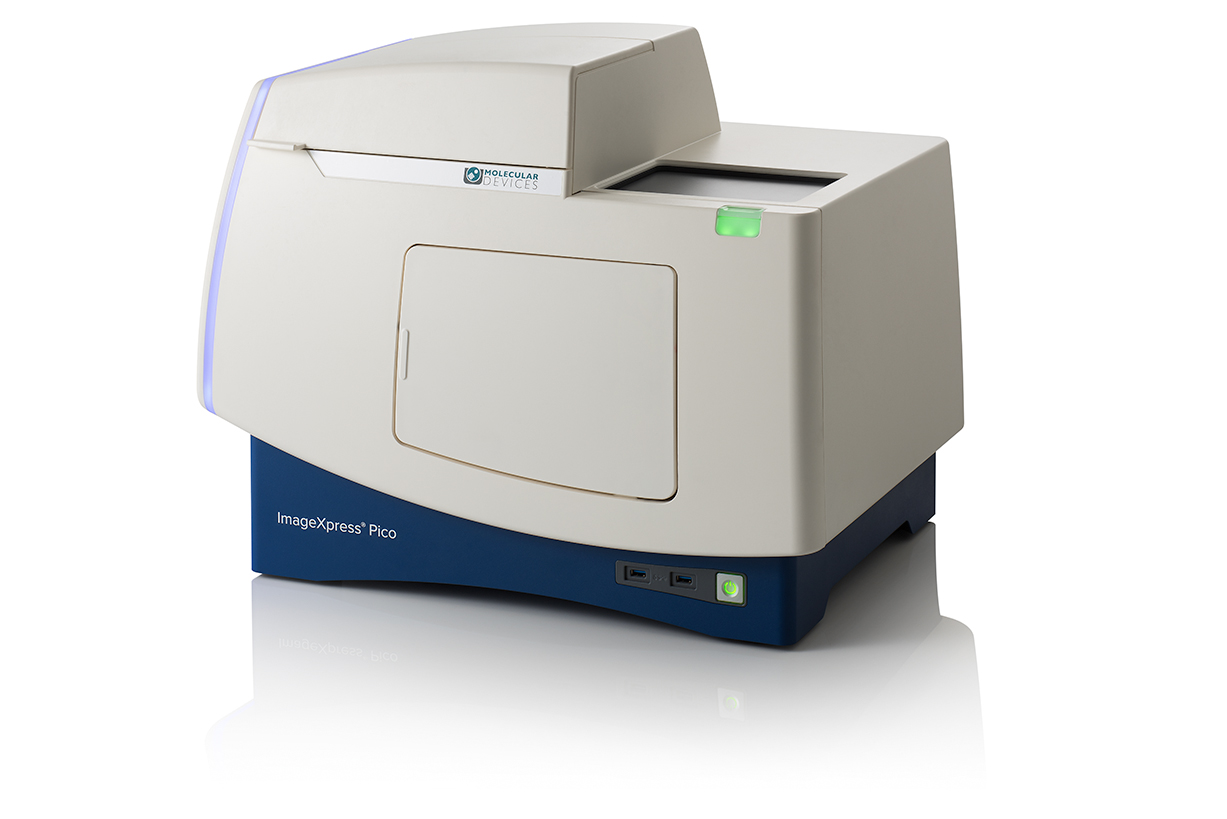
Automated fluorescence imaging that’s easier than digital microscopy

Get started quickly
With the approachable, icon-driven CellReporterXpress software, everyone can use the ImageXpress Pico system with minimal training to capture and analyze your first image.

Do more than cell counting
Expand your assays beyond cell counting with preconfigured templates for many commonly used cell imaging and analysis protocols such as apoptosis and detection of mitochondria.

Image economically
Alleviate the hassle of going to the core lab to run your samples. The system’s lab-friendly price affords the convenience of automated imaging and analysis on your lab bench.
Features

Multiple imaging modes
The ImageXpress Pico system provides user- and software-selectable objectives ranging from 4X to 63X, fluorescence imaging, live cell imaging, and brightfield imaging modes.

Predefined cell imaging protocols
The system offers preconfigured cell imaging protocols ranging from simple cell count to sophisticated neurite tracing analysis, removing the guesswork from optimizing parameters.

Plate-to-individual cell view
Data can be visualized at multiple levels from plate overviews to individual cells. Images are clickable to locate cells that match specific criteria.

Integrated data visualization tools
Liberate your lab from tedious digital microscopy, the software transforms images into data on the fly. Scatter plots, videos, and more can be generated automatically with just a few clicks.

Advanced slide scanning workflow
Whole slide scanning, for the determination of regions of interest, is quick and easy. Acquires higher resolution scans of only the select regions, saving time and data storage.

Temperature control
Temperature control up to 40◦C is standard on every ImageXpress Pico system, making live cell imaging a possibility.
General specifications
5 megaPixel CMOS Camera
Field of View with 10x: 1.93 mm2
4x-63X Air objectives
5 position filter cube changer
LED + image autofocus
Transmitted Light
Brightfield
Z-stack option
Digital confocal option
Up to 384-well plates & Microscope slides
Temperature control
Widefield
CellReporterXpress Analysis Software
Applications of ImageXpress Pico Automated Cell Imaging System
The CellReporterXpress Automated Image Acquisition and Analysis Software works with the ImageXpress® Pico systems. It has a clean, easy-to-learn interface for performing quantitative analysis on images acquired from automated microscopy. The software enables distributed analysis of images for increased throughput and is ideal for scaling microscopy imaging with slides or microplates. An icon-driven, linear workflow with a range of predefined protocols provides a streamlined user experience.

Analyze during acquisition
Reduce the time to run experiments with our integrated on-the-fly image analysis capability, allowing you to view numeric data during acquisition.

Simplify image analysis
Predefined analysis protocols for label-free and fluorescence imaging provides you with the ability to quickly run common biological assays while still allowing you to create and save your own protocols.

Share and access data from anywhere
The software makes sharing, presenting, and collaborating with peers easier, allowing multiple you to utilize the software simultaneously through browser-based remote access.
Features

Region selection for acquisition
Multiple unique positions within a well or slide can be selected to acquire and analyze only key areas of interest.

Label-free analysis
Automatically identifies objects in brightfield including small round cells, large cells, and beads.

Click-to-find feature
This feature auto calculates object parameters based on user-selected cells of interest, simplifying analysis setup for cell counting and for cellular responses.

Side-by-side cell magnification
Zoom or pan high-resolution images from whole well view down to cell level view. Simultaneously compare images from two wells.

Touch screen capability
Exceptional usability and precise control is built-in whether using a personal computer or a tablet.

Browser-based software
The ImageXpress Pico system can be controlled through a browser with data from any computer on the user’s network via Chrome™ or Safari®.
-
Apoptosis Analysis
Apoptosis is a process of programmed cell death important to a number of biological processes including embryonic development and normal tissue maintenance. Disruption in the regulation of apoptosis has been implicated in various diseases including cancer. Biochemical events lead to characteristic changes in cell morphology such as cell shrinkage, nuclear fragmentation, chromatin condensation, and mRNA decay, and ultimately cell death. Apoptosis can be initiated via the intrinsic or extrinsic pathways, and both pathways induce cell death by activating caspase enzymes

-
Cell Counting
Cell counting is fundamental and critical to numerous biological experiments. Assays such as drug compound toxicity, cell proliferation, and inhibition of cell division have a need to assess the number or density of cells in a well. Automated imaging can greatly speed up the cell counting process while reducing manual labor and human errors. Cells can be counted using a variety of methods, such as label-free cell counting under transmitted light or detection of nuclear dye with fluorescent imaging.

 ImageXpress Pico ismertető