


CloneSelect Imager
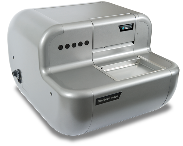
The CloneSelect™ Imager is a high-throughput automated solution for imaging and analyzing mammalian cells. Tracking the formation of a colony from a single cell is effortless as barcoded plates are tracked over time. Automated acquisition and analysis provide accurate, objective, and consistent results. Detailed imaging and comprehensive data analysis enable better decision making. With the ability to image a 96-well plate in 90 seconds, the imager will increase throughput and productivity.

Verify monoclonality easily
The Monoclonality Report feature streamlines the creation of supporting documentation for regulatory agencies. Reports are automatically generated based on parameters you select.

Detect cells accurately
Algorithms are optimized for accurate cell detection and address varying cell types and conditions. High resolution imaging provides accuracy and assurance of monoclonality.

Screen more clones in less time
The imager delivers industry leading acquisition times, allowing for imaging a 96-well plate in as little at 90 seconds.
Features

Choice of imaging modes
Label-free brightfield provides rapid acquisition without the use of harmful fluorescent agents. Customizable fluorescence imaging provides additional confidence of monoclonality.

Autogenerated data and tools
The software automatically calculates confluence measurements and generates growth curves, heatmaps, and image montages. Measurements for every well are automatically tracked over time.

Variety of plate formats and cell types
The imager is compatible with 6- to 384-well plates and enables use on suspension and adherent cell lines such as CHO, HEK, hybridomas, Per.C6®, Jurkat, SupT1, H-4-II-E, MCF-7, JT29, DLD1, and KB31.

Intelligent analysis
The software automatically calculates confluence for each imaging time point. Growth curve, image montage, total growth, and mean rate are generated automatically and are exportable.

Clear, crisp images
The imager features 4x objective lens with autofocus. High-resolution CCD camera images 1.85 microns per pixel, enabling accurate cell detection even at challenging areas such as well edges.

Custom automation options*
The Automated Workflow Engineering Solutions Team offer a variety of custom services from robotic plate loading to fully automated workstations with liquid handling and incubation.
*Price, time to deliver and specifications will vary based on mutually agreed technical requirements. Solution requirements may cause adjustment to standard performance.

CloneSelect Imager Monoclonality Report Feature
Providing image evidence of monoclonality in the cell line development process is not as simple as exporting an image of a single cell. For example, high-resolution images of the entire well…
Imaging
Software
Dedicated imaging software pre-installed on a high-specification PC, Microsoft Windows 10
White light imaging
Trans-illumination
Data tracking
1 x internal barcode reader for data tracking of each plate
Camera
High-resolution CCD camera
Imaging speed
<90 sec in standard mode
Resolution
Standard: 3.6 micron
Objective
4x
Instrumentation
Source plate capacity
1 x plates
Source plate type
Range of 6-, 12-, 24-, 96- and 384-well SBS microplates
Instrument dimensions
720 mm (width) x 428 mm (height) x 575 mm (depth)
Instrument weight
45 kg
Regulatory Approval
Compliance
CE
Quality
ISO9001:2008 certified
Applications of CloneSelect Imager
-

Cell Line Development
Cell line development is a critical step in the process of generating biopharmaceutical molecules, such as monoclonal antibodies. The process often begins with transfecting the host cell type with the DNA encoding the therapeutic protein of interest allowing for random or directed integration of target DNA into the host cell genome. Thousands of clones are screened to isolate the rare high producing cells, a manual and time-consuming process.
-

Cell Viability
Cell line development requires the discovery of single cell-derived clones that produce high and consistent levels of the target therapeutic protein. A critical first step in the process is the isolation of single, viable cells. Single-cells proliferate to form colonies, that can then be assessed for productivity of the target therapeutic protein. Viability and growth rates of single cell-derived clones can be characterized on the CloneSelect Imager.
-

Monoclonality Assurance
Cell line development and assurance of monoclonality are critical steps in the process of generating biopharmaceutical molecules, such as monoclonal antibodies. A cell line can be established following the isolation of a single viable cell robustly expressing the protein of interest. A key milestone in this process is documenting evidence of clonality. Documentation of clonality is typically image-based, whereby an image of a single cell is produced and included in regulatory filings.
-

Label-Free Cell Migration
Wound healing assays are used in a range of disciplines to study cell migration, the coordinated movement of a cell population. A manual or standardized wound is generated on a monolayer of cells grown in microplates. The microplates can be screened against a library to study the effect on cell migration. The effect of a library of compounds or genes can be studied using this approach. The CloneSelect Imager enables the rapid imaging (
-

Colony Forming Assay
The soft agar colony formation assay is a classical and renowned technique for characterizing the ability of cells to undergo anchorage-independent growth, a hallmark of carcinogenesis. After seeding in a matrix that enhances colony formation, such as a semi-solid media, cells are typically incubated with compounds that may affect colony growth. The CloneSelect Imager can image every well and automatically calculate confluence, estimate colony number and area, and track colony growth.