


CytoFLEX platform
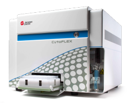
New standards of fluorescence sensitivity <30 MESF-FITC, <10 MESF-PE
Violet side scatter resolution for particle detection < 0.2µm
Quality performance, multiple configurations for real power on your benchtop
The high-performance CytoFLEX system offers scalable detection capabilities for up-to three lasers and 13-color research flow cytometry right on your benchtop.
Ready to run in minutes, not hours with CytoFLEX
The CytoFLEX system is easy to learn, easy to operate and easy to maintain.
- Superior sensitivity and resolution for excellent fluorescence and nanoparticle detection
- Compact, reliable design fits right on your benchtop
- Future-proof system with easy upgrade options
- Powerful performance with up-to 21 flexible configurations (up-to three Lasers, 13 colors)
- CytExpert experiment-based software for easy multicolor setup and QC
- Versatile plug-n-play filters (bandpass-only light collection)
- Integrated optics for alignment stability
- Accommodates 12x75mm and microtiter tubes
Sensitivity
An intelligent patent-pending optical design optimizes excitation from up-to three lasers (488nm; 638nm; 405nm) and light collection efficiency. With unique flow cell design and integrated optics, the innovative Wavelength Division Multiplexing (WDM) detection module includes solid-state, high efficiency, low-noise detectors for excellent performance. The WDM’s bandpass-only design guarantees flexibility with easily changeable filters and 4-13 configurations.

Nano particle detection
When equipped with a violet laser, the CytoFLEX can be set up to measure Side Scatter from 405nm for enhanced nanoparticle detection. Mie theory predicts that the scattering cross section of a particle, and thus its scattering intensity, depends on the wavelength of light, the angle of collection, and the size, shape, and refractive index of the particle. All other factors being equal, using a shorter illumination wavelength will result in an increase in scattering cross section, and thus more scattered light. The CytoFLEX offers light scattering parameter measurements from the 488nm laser light, and optionally from the 405nm laser to enhance small particle detection.

Maximize investment in CytoFLEX with custom configuration options
With future-proof upgrade options for up-to 13 colors, the CytoFLEX system evolves as your application needs change. Upgrade your configuration in less than one day knowing that all CytoFLEX instruments have been fully tested across all configuration types.
| Excitation | Fluorescence Channels | Fluorochromes |
|---|---|---|
| 488nm | 525/40 BP | FITC, Alexa Fluor 488, CFSE, Fluo-3 |
| 585/42 BP | PE, PI | |
| 610/20 BP | ECD, PE-Texas Red, PE-CF594, PI | |
| 690/50 BP | PC5, PC5.5, PerCP, PerCP-Cy5.5, PI | |
| 780/60 BP | PC7 | |
| 638nm | 660/10 BP | APC, Alex Fluor 647, eFluor 660 |
| 712/25 BP | APC-Alexa Fluor 700, Alexa Fluor 700 | |
| 780/60 BP | APC-Alexa Fluor 750, APC-Cy7, APC-H7, APC-eFlour 780 | |
| 405nm | 450/45 BP | Pacific Blue, V450, eFluor 450, BV421 |
| 525/40 BP | Krome Orange, AmCyan, V500, BV510 | |
| 610/20 BP | Violet610, BV605, Qdot 605 | |
| 660/10 BP | Violet660, BV650, Qdot 655 | |
| 780/60 BP | Viole780, BV785, Qdot 800 |
CytoFLEX Optics
Excitation optics
The instrument has the capacity for 15 parameters, including 13 for fluorescence detection. The fully activated instrument includes five channels from the 488 nm (Blue) laser, three from the 638 nm (Red) laser, and five from the 405 nm (Violet) laser. Instruments with as few as four fluorescent channels activated are available with the ability to activate additional parameters as needed by purchasing an activation key.
Flow cell
Fixed integrated optics and quartz flow cell design with >1.3 numerical aperture.
Flow Cell dimensions: 420 μm x 180 μm internal diameter
Forward scatter detection
Proprietary Axial Light Loss (ALL) sensor system using silicon photodiodes with built in 488/8 μm band pass filter.
Fluorescence and side scatter detection
Fluorescence and side scatter light delivered by fiber optics to Avalanche Photo Diode detector arrays. Proprietary design ensures high performance, high efficiency, low-noise signal detection. Emission profiles are collected using reflective optics and single transmission band pass filters.
Violet side scatter configuration
Option to configure Avalanche Photo Diode detector array to collect side scatter signal from Violet (405 nm) laser. The configured channel (VSSC) can be utilized to better resolve nanoparticles below 200 nm size threshold.
Fluidics
Ultra-low pressure peristaltic sheath and sample delivery system
- Low maintenance system
- Sheath Fluid Filter and Sample Pump Tubing can be replaced by the user (no service visit required)
Sample flow rates
- Fixed Flow Rates: 10, 30 and 60 μL/min.
- Custom Flow Rate Control mode from 10 to 240 μL/min in 1 μL increments.
- Gravimetric calibration for absolute counts within CytExpert Software.
Fluid capacity
- Standard 4 L tanks
- Optional 10L cubitainers
Automated maintenance functions
- System Startup, Sample Mixing, Backflush, Prime, Shutdown, Deep Clean
Sample input format
- 5 mL (12 x 75 mm) polystyrene and polypropylene tubes
- 1.5 mL and 2 mL microcentrifuge tubes
Plate loader formats
- 96-well Standard Flat, U and V bottom plates
The Unique Peristaltic Sample Delivery System of the CytoFLEX Analyzer Enables Optimized Measurements
Generation of fluorescent antibody or genetic labels to identify hormone and neurotransmitter receptoractivity can be difficult and time consuming. A useful alternative is recording physiological changes inresponse to agonist binding its cognate receptor, many of which are G-protein coupled. When an agonist binds a G-protein coupled receptor, it triggers a quick cascade of events that often results in a transient release of calcium from intracellular stores. Alterations in transient intracellular calcium ([Ca2+]i) levels have been used previously in flow cytometry to identify functional receptor expression in cellular subpopulations [2], here we show that peristaltic sample delivery of the new CytoFLEX analyzer is particularly well suited to agonist-based calcium studies. Using the ester based, green fluorescent calcium indicator, Fluo-4 AM (Life Technologies), [Ca2+]i changes were measured in HEK-293 cells in response to ATP stimulation. Simple plumbing modifications to the CytoFLEX allowed easier access to the sample tube for agonist application; further modifications were made to implement a “stop time” technique. By using response to agonist as our physiological criteria, we have fundamentally enabled receptor identification and conclusively demonstrated its functionality.
CytoFLEX Instrument Evaluation Using Biological Specimens
Evaluation of the Xitogen flow cytometer, by independent research flow cytometry laboratories and Beckman Coulter senior research and development scientists, was performed using flow cytometers equipped with 12 parameters and 9 fluorescent detectors. Instruments were equipped with the enhanced Side Scatter channel (VSSC) option. Evaluation of biological specimens included fresh human whole blood, human leukocyte control cells, and smaller cells and particles, such as RBCs, platelets, microbes, liposomes, and vesicles.
Set-Up Of The CytoFLEX For Extracellular Vesicle Measurement
The measurement and the characterization of Extracellular Vesicles (EV) have been of growing interest over the last 20 years. Flow cytometry instruments were not the most appropriate way to analyse these particles as the optical resolution of instruments was insufficient to detect particles below 250nm. However, the Beckman Coulter CytoFLEX* now offers the ability to measure EV down to at least 150nm and allows the detection of their cellular origin using up to 13 fluorescence parameters. Regardless of the technical improvements the set-up of the instrument is still a critical point and several requirements need to be met which are illustrated here.
Extracellular vesicles are a heterogeneous cell-derived particle population in a size range between 50nm to 1,000nm. There is a growing interest not only from academic research groups to determine EV in several fluids such as cell culture supernatant, in plasma samples or in whole blood but also in clinical research since it has been shown that the measurement of microparticles (MPs)1 might be of clinical relevance. The methods to identify EV are many and involve high speed centrifugation, Western blotting, proteomics, electron microscopy, imaging methods and flow cytometry. Methods for the detection of EV by flow cytometry have been developed in the last years and special attention has been paid to standardization protocols. Compared
with other methods, flow cytometry has the big advantage that EV can be detected as rare events, in high numbers and by antigens on the surface, which characterize their cellular origin.
Andreas Spittler, MD, Associate Professor for Pathophysiology, Medical University of
Vienna, Core Facility Flow Cytometry & Department of Surgery, Research Laboratories