


ScanLater Western Blot Detection Cartridge
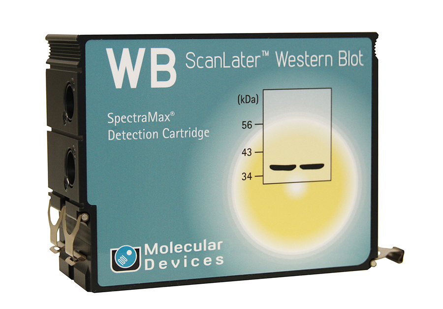
Transform your SpectraMax® i3 or Paradigm® System into a western blot readerThere are often a great many western blots along the path between imagination and understanding. Now you can read them using the advanced optical technology that's already in your lab — inside your microplate reader. The ScanLater™ Western Blot Detection System includes the ScanLater Western Blot Detection Cartridge, ScanLater Western Blot Assay Kit, and image acquisition powered by SoftMax® Pro Software. In just minutes, you can install the ScanLater Western Blot Detection Cartridge in a SpectraMax® i3 or SpectraMax® Paradigm® System to add western blot detection capability, rather than buying a dedicated western blot detection system. See more, and do more, with your microplate reader.
The ScanLater Western Blot Detection Cartridge is a user-installable option for the SpectraMax i3 and SpectraMax Paradigm Multi-Mode Platforms. Leveraging this novel technology, researchers can:
- Add western blot detection to their microplate reader within 2 minutes
- Eliminate time-dependent substrate addition steps
- Maintain femtogram to picogram protein sensitivity comparable to traditional western blot methods
- Sustain blot signal stability for up to 30 days
- Use a single software platform SoftMax Pro Software, to run both microplate and western blot detection assays
Features:
- User-installable cartridge for the SpectraMax i3 and Paradigm Multi-Mode Platforms
- Time-resolved fluorescence detection
- Xenon flash lamp excitation source
- 340/80 nm excitation range
- 616/10 nm emission range
- Supports nitrocellulose and PVDF membranes
- Compatible with goat anti-rabbit, anti-mouse or streptavidin labeled antibodies
- Reads membranes fitting the ScanLater SpectraMax Membrane Holder
Technologies of Western Blot Cartridge

-
Schematic representation of emission lifetimes that minimize background fluorescence
Schematic representation of emission lifetimes of background fluorescence and Eu-chelate showing principle of TRF measurement. Signal is measured after appropriate time-delay to reduce auto-fluorescence.
-

ScanLater Western Blot Assay Workflow
-

ScanLater Western Blot Assay Principle
Schematic of protein detection method shows the secondary antibody is directly labeled with Europium.
-
 Europium-Chelate Excitation and Emission Spectra
Schematic representation of Europium-chelate excitation and emission spectra
Representative applications:
- Protein identification and quantitation
- Epitope mapping
- Amino acid composition and sequence analysis
- Spots imprinting
- Phosphoprotein endogenous or exogenous expression
- Protein resillience
- Structure domain analysis
- Protein expression
- Protein content in serum
- QC elimination of albumin and lgG in serum
- Protein expression the cell cycle
| Product Name | Description | Part Number |
| System ScanLater Western Blot Detection System |
|
0200-7027 |
| Assay Kit ScanLater: Eu-Streptavidin Evaluation Kit |
|
R8200
|
| Assay Kit ScanLater: Eu-Streptavidin Bulk Kit |
|
R8203
|
| Assay Kit ScanLater: Anti-Mouse Evaluation Kit |
|
R8201
|
| Assay Kit ScanLater: Anti-Mouse Bulk Kit |
|
R8205
|
| Assay Kit ScanLater: Anti-Rabbit Evaluation Kit |
|
R8202
|
| Assay Kit ScanLater: Anti-Rabbit Bulk Kit |
|
R8204
|
| Assay Kit ScanLater: Anti-Goat Evaluation Kit |
|
R8224
|
| Assay Kit ScanLater: Anti-Goat Bulk Kit |
|
R8227
|
| Assay Kit ScanLater: Anti-Rat Evaluation Kit |
|
R8225
|
| Assay Kit ScanLater: Anti-Rat Bulk Kit |
|
R8228
|
| Protein Ladder ScanLater Western Blot Protein Ladder |
|
R8220
|
| Assay Kit Components | ||
| ScanLater Wash Buffer (100 mL) | R8206 | |
| ScanLater 5x Blocking Buffer (120 mL) | R8207 | |
| ScanLater Goat Anti-Mouse Eu-labeled Antibody (60 µg) | R8208 | |
| ScanLater Goat Anti-Rabbit Eu-labeled Antibody (60 µg) | R8209 | |
| ScanLater Eu-labeled Antibody Streptavidin (60 µg) | R8212 | |
| ScanLater Eu-Anti-Goat IgG (60 μg) | R8226 | |
Related Video
Data Sheet
Application
- Detection and Quantitation of Protein with ScanLater Western Blot Detection System
Poster
- Western Blot Protein Detection and Quantitation Based on Europium Labeled Proteins Using a Plate Reader